Corals in 3D: Using laser scanning to capture complex coral morphologies
Corals in 3D: Using laser scanning to capture complex coral morphologies
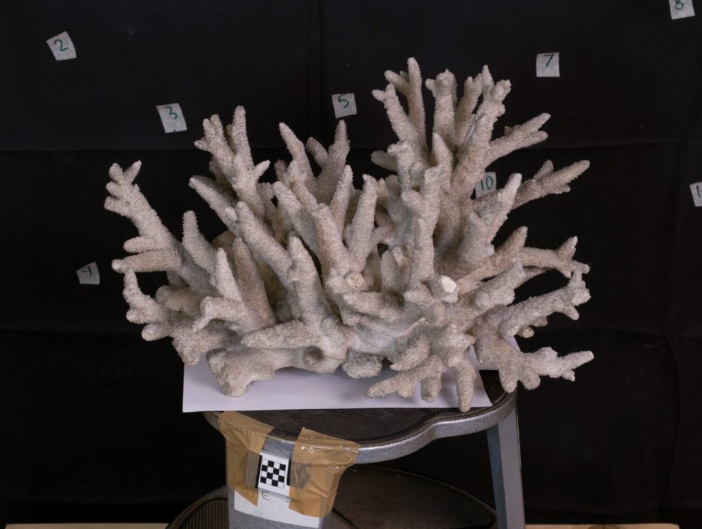
The form that corals take when they grow has multiple consequences for their biology and ecology, and has wider implications for the ecology of other reef taxa. The variation in shape that corals can have ranges from simple, boulder-like forms all the way up to intricate interwoven networks of branches. Despite its importance however, fully capturing the shape of a coral colony has until recently proven difficult for the more complex morphologies. An ongoing project by Kyle Zawada under the supervision of Maria Dornelas and Josh Madin, has proven successful in capturing the shapes of corals that span many growth forms and sizes via high precision laser scanning equipment provided by CREAFORM free of charge via their studentship programme.
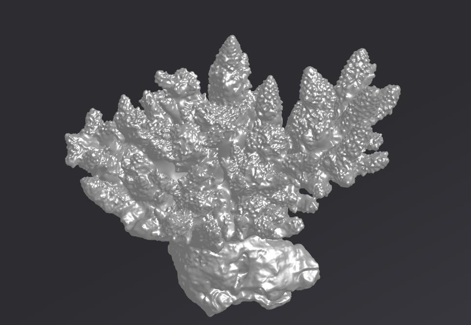
By working with the large collections of coral skeletons available at the Natural History Museum in London with Miranda Lowe and the Museum of Tropical Queensland in Townsville with Tom Bridge, a catalogue of over 230 coral specimens have now been digitised ready for data analysis. The team hopes that these meshes will be able to provide a detailed picture of coral morphology, and will allow them to explore the consequences of shape on the biology and ecology of corals in a more in depth way than was possible beforehand.